
The jaw, chewing muscles, and posture are interconnected. This article describes how we systematically examine the temporomandibular joints and chewing muscles before planning treatment – to ensure the underlying cause of symptoms is not overlooked.
Structured functional diagnostics of the temporomandibular and musculoskeletal system as the basis for treatment planning.
Part 1 of the article series: Brief screening and ongoing functional diagnostics before every orthodontic treatment
This article is part of a three-part series in the Journal of Aligner Orthodontics. Part 1: How the temporomandibular joint, muscles, and posture are examined (this page) · Part 2: How the relaxed jaw position is determined · Part 3: What happens after the splint
Why Thorough Diagnostics Extend Beyond the Teeth
Before orthodontic treatment begins, one question must be clarified: Are the temporomandibular joints in a relaxed, healthy position — or does the bite force the lower jaw into an unfavorable position? If an unrecognized dysfunction of the temporomandibular joint (medical term: temporomandibular disorder, abbreviated TMD) is overlooked, it can jeopardize the success of the entire treatment. This publication describes how such a dysfunction is systematically identified — and why not only the teeth but the entire body plays a role.
The reason: The temporomandibular joint is closely connected to the neck and back musculature and to posture via muscle and fascial chains. A dysfunction in the jaw can extend into the neck, shoulders, and back – and vice versa.1 Pain often does not occur where the cause lies. For this reason, modern functional diagnostics considers the craniomandibular system (temporomandibular joint and masticatory muscles) together with the musculoskeletal system (posture and movement apparatus).
The First Step: Brief Screening
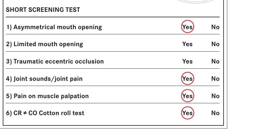
At the beginning is a brief screening test2that makes sense before treatment for every patient – for adults as well as in orthodontics for children and adolescents. Six points are examined:
- asymmetric mouth opening,
- restricted mouth opening,
- abnormal (“traumatic”) bite,
- joint sounds or joint pain,
- pain when palpating the chewing muscles,
- a difference between the habitual bite and the relaxed jaw position (so-called cotton roll test).
If two or more of these points are abnormal, comprehensive functional diagnostics follow immediately.
Every point in the brief screening is answered with yes or no. The authors developed the examination form together with physicians working in manual therapy; the results are transferred directly into the detailed form. In the example from the publication, a case from our practice, four out of six points were abnormal—making the comprehensive examination necessary. You can get a first impression in advance with our TMD self-test ; it does not replace the examination.
Comprehensive Examination of the Temporomandibular Joint and Chewing Muscles
In the in-depth diagnostics, several areas are systematically assessed and documented on a standardized examination form:
Mobility of the Lower Jaw
The maximum mouth opening (the distance between the incisal edges), lateral movements, and protrusion of the lower jaw are measured. The movement pattern during opening is also important: Does the lower jaw deviate to the side and return (deviation), or does it remain deflected (deflection)? Such patterns provide clues about the position of the articular disc (meniscus).3
Palpation of the Temporomandibular Joints
The joints are gently palpated both externally (in front of the ear) and through the ear canal. This allows for comparative assessment of joint sounds such as clicking or grinding, pressure pain, swelling, and mobility of the condyles on both sides. Palpation through the ear canal in particular permits very precise evaluation of the sensitive area behind the condyle.
Examination of the Muscles
Masticatory, temporal, neck, and shoulder muscles are specifically palpated. Particular attention is given to so-called trigger points – small, tense muscle areas that can radiate pain to other regions.4 They are treated first, as they often significantly shape the clinical presentation.
Bite in different positions
The tooth contacts in the habitual bite are compared with those in a more relaxed, physiological jaw position—determined using the simple cotton roll test. If there is a clear difference, this is an important clue: every treatment should be oriented to the relaxed joint position, not the forced habitual bite.
Looking at the whole body
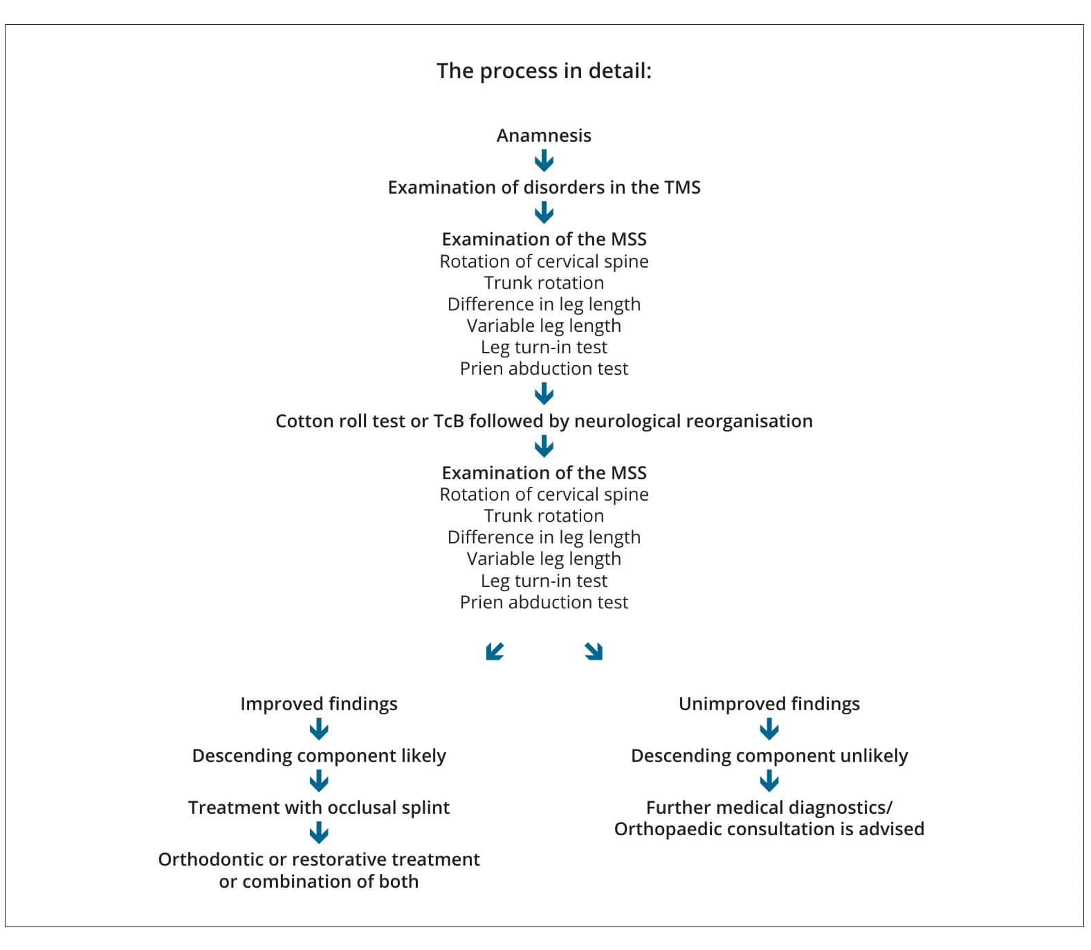
Next, the postural and movement apparatus is included. Among other things, the rotation of the cervical and thoracic spine, differences in leg length, and special manual tests (e.g., the Prien hip abduction test)5are examined. Facial symmetry is also assessed, as a lack of support in the posterior tooth region on one side can be reflected in slight facial asymmetry.6
Then follows the cotton-roll test: Cotton rolls are placed between the first small molars for about two to three minutes so that the teeth no longer engage in the habitual bite. Afterward, you swallow once and take a few steps; then the same body tests are repeated. Do the findings improve? If yes, this suggests that the bite is indeed involved in the complaints in the neck, back, or hips—what is called a “descending” chain. If nothing improves, the cause is more likely elsewhere, and medical or orthopedic evaluation is advisable.
From diagnosis to treatment
Only when this complete picture is available can a meaningful decision be made about how to proceed. If the medical history and examination indicate a treatable functional disorder, the exact, relaxed jaw position is first determined—how this is done is described in the article on determining the therapeutic jaw position. This is often followed by preliminary treatment with an occlusal splint that can be removed at any time. If a cone-beam CT or MRI is needed to clarify a joint finding in more detail, we will discuss this with you beforehand. After successful follow-up treatment , the jaw position can be stabilized permanently.
The sequence is important: first understand, then treat. Orthodontic correction that begins without this diagnostic work runs the risk of cementing an unfavorable jaw position rather than resolving it.
What to Expect at Your Examination Appointment
After booking the appointment, you will receive a link by text message or email to our digital medical history form “Nelly”; there you can enter your symptoms, previous illnesses, and medications from home. At the practice, we measure the mouth opening, palpate the temporomandibular joints and muscles, and examine the cervical spine, leg length, and hips while you are lying down and sitting. Clinical and instrumental functional analysis as well as temporomandibular joint measurement with the DMD system are not covered by statutory health insurance; they are billed privately according to the German Fee Schedule for Dentists (GOZ). What applies in your case depends on the scope of the examination—you will receive the amount in writing before the examination.
Whether you have statutory or private health insurance: diagnostics, consultation, and care are the same for all patients at our practice.
What systematic diagnostics means for treatment
Thorough functional diagnostics are the foundation of any successful treatment for temporomandibular joint disorders. They combine the precise examination of the temporomandibular joint and masticatory muscles with a view of posture and the musculoskeletal system—and thus make visible what a simple examination of the teeth would overlook. Based on this, we plan further treatment and also tell you if the cause is more likely to be outside the jaw.
Sources
- Rocabado, M. (1983). Biomechanical relationship of the cranial, cervical, and hyoid regions. Journal of Craniomandibular Practice, 1(3), 61–66. https://doi.org/10.1080/07345410.1983.11677834
- Meyer, G. (2018). Short clinical screening procedure for initial diagnosis of temporomandibular disorders. Journal of Aligner Orthodontics, 2(2), 91–98.
- Bumann, A., & Lotzmann, U. (Hrsg.). (2000). Funktionsdiagnostik und Therapieprinzipien. Thieme.
- Travell, J. G., & Simons, D. G. (Hrsg.). Handbuch der Muskeltriggerpunkte: Obere Extremität, Kopf und Thorax. Elsevier, Urban & Fischer.
- Schupp, W., Boisserée, W., Haubrich, J., Hermens, E., Grunert, I., Rottler, A., & Meyer, U. (2015). Diagnostische Verfahren im kraniomandibulären System. Manuelle Medizin, 53(1), 47–59. https://doi.org/10.1007/s00337-014-1174-5
- Schupp, W., Oraki, A., Haubrich, J., Freesmeyer, W., & Kopsahilis, N. (2009). Okklusionsveränderungen und deren Auswirkungen auf den Halte- und Stützapparat. Manuelle Medizin, 47(2), 107–111. https://doi.org/10.1007/s00337-009-0673-2
Your orthodontist in Cologne!
Schedule your no-obligation consultation at JUST KFO – Dr. Steinmaier & Kollegen in Cologne.
