Before fabricating a splint, the relaxed rest position of the mandible must be determined. This article explains how to identify this position and distinguish it from the habitual bite.
Determination of rest position and construction bite versus habitual bite.
Part 2 of the article series: the relaxed (centric) position of the temporomandibular joints and the therapeutic construction bite compared to the centric bite
This article is part of a three-part series in the Journal of Aligner Orthodontics. Part 1: How the temporomandibular joint, muscles and posture are examined · Part 2: How the relaxed jaw position is determined (this page) · Part 3: What happens after the splint
The Correct Jaw Position – Why It Matters So Much
In most joints of the body, muscles determine where the joint sits. The temporomandibular joint is different: as soon as the teeth bite firmly together, they dictate – through the occlusion – the final position of the joint.1 If this habitual bite does not match the relaxed, healthy joint position, it can cause problems over time. For any treatment of temporomandibular joint problems, a healthy, relaxed jaw position must first be found. That is exactly what this professional publication is about.
This relaxed position – in technical terms “centric” or physiological jaw position – is not a fixed value, but varies from person to person and can change slightly depending on muscle tension, posture and daily condition.2 There is no single “true” measurement method for it; rather, the goal is to find a position that fits the entire jaw and musculoskeletal system. This determination is always preceded by a thorough3 functional diagnostic evaluation.
Centric Bite and Therapeutic Construction Bite
To record the jaw position, a bite registration is taken – in simple terms, a precise impression of the bite position in a wax plate. Two situations are distinguished:
Centric Bite in Healthy Joints
For joints without symptoms, a relatively simple centric bite is sufficient. For this, a 3 millimeter thick wax plate is heated in a water bath to 52 °C, cut into a trapezoidal shape and fitted to the upper teeth. The patient bites into it in an upright, relaxed posture – without the treating orthodontist guiding the lower jaw. The wax is then hardened in ice water. Afterwards, the contact points are carefully checked and reduced until a stable, reproducible bite position is achieved.
Therapeutic Construction Bite in Compromised Joints
For symptoms or diseased joints, that is not enough. Here, a therapeutic construction bite is taken – while simultaneously palpating the joints through the ear canal. This allows mobility, symmetry and the sensitive area behind the condyle to be precisely assessed while the correct position is adjusted. Before taking the bite, the joints are gently mobilized and muscle tension is released; accompanying manual therapy is ideal.4, 7 A particularly hard wax is used that remains dimensionally stable in the mouth and thus allows manual control.
From your habitual bite, you shift your lower jaw forward about 2 millimeters, with light tooth contact. Then the treating orthodontist takes over the guidance and palpates the condyles through the ear canal with the fingertips until the joints are free and without pressure. As a patient, you primarily feel the palpation at the ear and the biting into the wax.
Full-Body Examination
Whether the identified position is truly correct is not only evident in the jaw. With the construction bite in place, the patient is asked to swallow once and take a few steps – this allows the body to reorganize itself. Subsequently, posture and mobility tests are repeated.5 The same tests are repeated as during the initial examination: rotation of the cervical and thoracic spine, leg length comparison, and hip tests. What these tests are is described in Part 1 of the series. If these findings improve significantly with the new bite, this is a strong indication that the jaw position truly fits the body and musculature. This comparison makes the determination more objective and reliable.
From Bite Registration to Appliance
The recorded position is transferred to an articulator using a facebow and the construction bite – a device that accurately replicates jaw movements outside the mouth.6 Only this precise model mounting allows reliable analysis of the occlusion and the fabrication of a precisely fitting occlusal splint. Without this careful transfer, the position would be inaccurate and unusable for both diagnosis and splint fabrication.
In the articulator, the bite is checked with a color foil only 12 micrometers thick. This makes even contacts visible that no one could detect in the mouth.
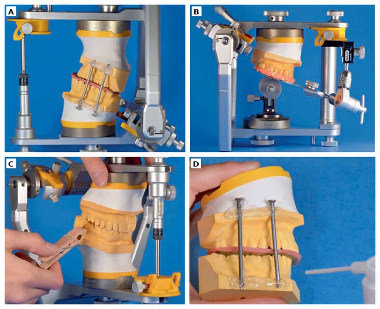
A case we treated in our practice and described in the publication shows why this is important. A patient came in because of severe back pain. In her mouth, her bite looked stable, the teeth were straight. Only the models mounted in the articulator in a relaxed jaw position showed: In this position, only the incisors touched, with a gap remaining at the canines and molars. When biting in the habitual manner, the lower jaw had to shift backward and upward. DVT and MRI images confirmed the retruded condyles and an anteriorly displaced articular disc.
In the authors’ view, treatment that only considers the teeth would have missed the cause here. They summarize this with a guiding principle they attribute in the publication to American dentist Harold Gelb (1994): “Think orthopaedic first – then teeth,” meaning first the joint and function, then the teeth.
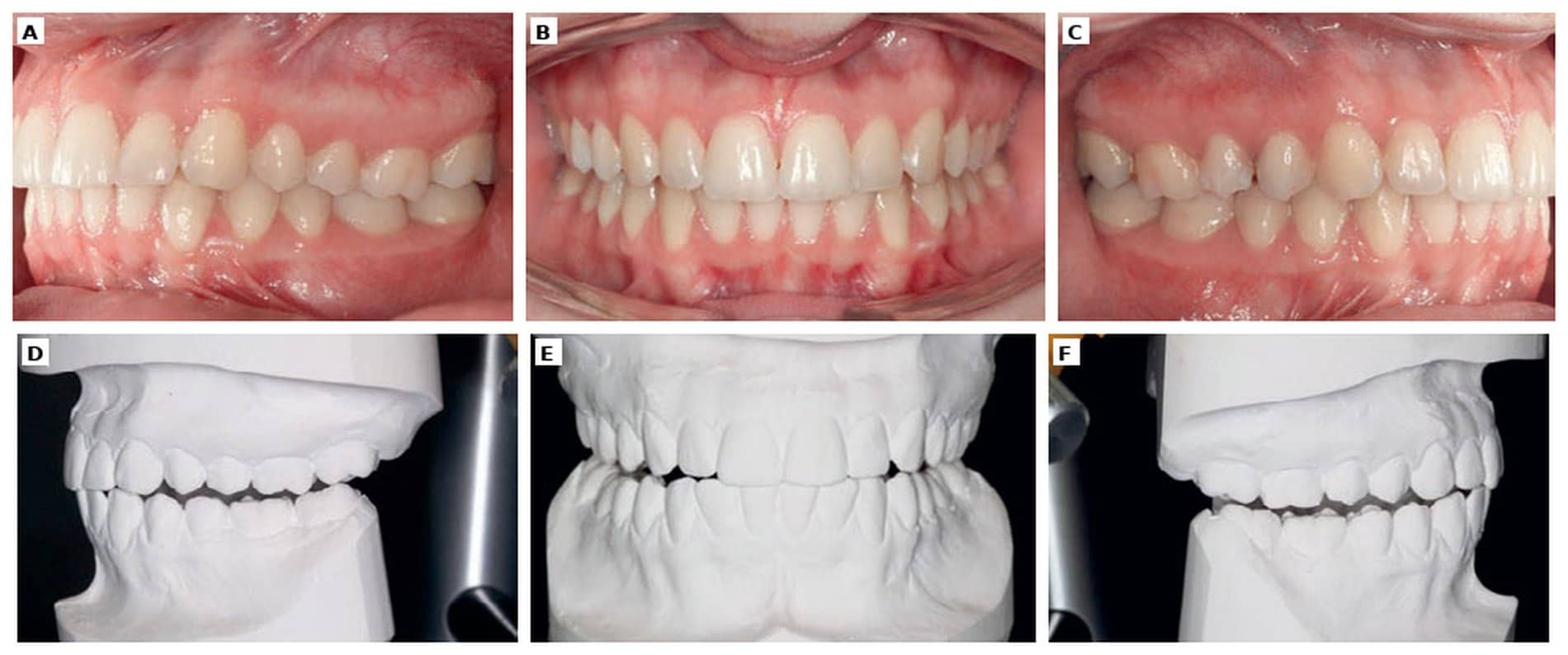
The patient was treated with a lower jaw splint, which she wore except during eating and brushing, combined with physiotherapy. In this case, she reported after five weeks that she no longer had back pain or temporomandibular joint pain. How quickly and how strongly symptoms improve varies from person to person. What happens after such a splint phase is described in Part 3 of the series.
Also Suitable for Children
The described procedure can also be applied to children and adolescents, for example when a construction bite is needed for a removable functional orthodontic appliance. Here too, simultaneous palpation of the joints provides valuable additional information.
Diagnosis and Therapy Belong Together
The relaxed, healthy jaw position is the foundation of any treatment for temporomandibular disorders. It cannot simply be “read off” but must be carefully worked out and verified across the whole body. A carefully taken construction bite, combined with palpation findings and model mounting in the articulator, is the basis for a precisely fitting splint and for all further treatment steps.
Does taking the bite hurt?
Palpating the joints can be uncomfortable if the joints are inflamed or irritated; this is also a finding that we note. Biting into the wax itself is not painful. If anything presses too hard, please say so immediately.
Why a wax bite when the practice has an intraoral scanner?
The scanner captures the teeth, but not the position in which the temporomandibular joints are relaxed. This position is palpated by hand and recorded in the wax. For splints that we fabricate digitally, this wax bite is then also scanned and transferred into the computer planning. For aligner planning, digital recording of jaw movements is added; how this works is described in our article on the virtual articulator.
Who covers the costs?
Clinical and instrumental functional analysis as well as temporomandibular joint measurement with the DMD system are not covered by statutory health insurance; they are billed privately according to the fee schedule for dentists (GOZ). Whether covered by statutory or private insurance: diagnostics, consultation, and care are the same for all patients at our practice. More under Costs & Insurance.
Publication discussed: Schupp, W., Funke, J., & Boisserée, W. (2018). Continuing diagnostics and therapy of the temporomandibular and musculoskeletal system (TMS/MSS): The rest position of the temporomandibular joint (TMJ) and the therapeutic construction bite vs. the centric bite. Journal of Aligner Orthodontics, 2(4), 267–281. (J. Funke = Dr. Julia Steinmaier)
Sources
- Kubein-Meesenburg, D. (1985). Die kraniale Grenzfunktion des stomatognathen Systems des Menschen. München: Hanser.
- Türp, J. C., Schindler, H. J., Rödiger, O., Smeekens, S., & Marinello, C. P. (2006). Vertikale und horizontale Kieferrelation in der rekonstruktiven Zahnmedizin. Schweizer Monatsschrift für Zahnmedizin, 116(4), 403–417.
- Meyer, G. (2018). Short clinical screening procedure for initial diagnosis of temporomandibular disorders. Journal of Aligner Orthodontics, 2(2), 91–98.
- Schupp, W., & Marx, G. (2002). Manuelle Behandlung der Kiefergelenke zur Therapie der kraniomandibulären Dysfunktionen. Manuelle Medizin, 40(3), 177–183. doi:10.1007/s00337-002-0133-8
- Schupp, W., Boisserée, W., Haubrich, J., Hermens, E., Grunert, I., Rottler, A., & Meyer, U. (2015). Diagnostische Verfahren im kraniomandibulären System. Manuelle Medizin, 53(1), 47–59. doi:10.1007/s00337-014-1174-5
- Meyer, G. (Hrsg.). (1993). Die physiologische Zentrik im Rahmen der instrumentellen Okklusionsdiagnostik. Funktionslehre. Hanser.
- Arribas-Pascual, M., Hernández-Hernández, S., Jiménez-Arranz, C., Grande-Alonso, M., Angulo-Díaz-Parreño, S., La Touche, R., & Paris-Alemany, A. (2023). Effects of Physiotherapy on Pain and Mouth Opening in Temporomandibular Disorders: An Umbrella and Mapping Systematic Review with Meta-Meta-Analysis. Journal of Clinical Medicine, 12(3), 788. doi:10.3390/jcm12030788
Your orthodontist in Cologne!
Schedule your no-obligation consultation at JUST KFO – Dr. Steinmaier & Kollegen in Cologne.
Book online 24/7 in a few clicks · free parking in our courtyard