
Occlusal splints for TMD can now be digitally planned and fabricated. This article describes the process from impression to finished splint.
Digital workflow for fabricating occlusal splints considering orthopedic principles.
The Occlusal Splint: The First Step in Treating Temporomandibular Disorders
When teeth are being straightened, the visible alignment is often the primary focus. However, it is equally important that the teeth meet correctly at the end and that the temporomandibular joints function harmoniously. This is precisely where the occlusal splint comes in. For temporomandibular joint disorders (temporomandibular disorder, or TMD), it is often the first, conservative treatment step — even before actual tooth correction.1 The underlying specialist publication describes this approach and the modern, digital fabrication of the splint. Below, we explain the key points in understandable terms.
What Is an Occlusal Splint?
An occlusal splint is a removable appliance made of rigid acrylic, usually worn in the lower jaw. It deliberately adjusts how the upper and lower jaws relate to each other. In professional terminology, it is also called a craniomandibular orthopedic repositioning appliance (COPA)2 because it guides the mandible and the condyles (the rounded ends of the jaw joint) into a relieved position. Its main purposes are the treatment of TMD and the protection of enamel and the periodontal apparatus.
Why the Splint First—and Only Then the Orthodontic Correction?
Ideally, every orthodontic treatment should be planned from a healthy, physiological position of the temporomandibular joints — professionally called “centric relation” (CR) — and not from the habitual bite position where the teeth currently fit together (the “habitual intercuspation,” CO).3 When a functional disorder is present, there is often a gap between these two positions. The occlusal splint is a reversible measure — meaning it is completely reversible — that first establishes and verifies this healthy joint position. Only when it is clear from which position treatment should proceed does the actual tooth correction begin.
How Is the Correct Position Determined?
It begins with a thorough functional analysis. This starts with a standardized short test (the “Short Screening Test”)4 and an examination of the interaction between the jaw, musculature, and body posture.5 The short test comprises six points: whether the mouth opens asymmetrically or with limited range, whether individual teeth are heavily loaded during lateral movements, whether the temporomandibular joints pop or hurt, whether the chewing muscles are sensitive to pressure, and whether the bite changes when biting briefly on two cotton rolls. This cotton roll test reveals whether the habitual bite position differs from the relaxed joint position. If two or more points are conspicuous, a comprehensive functional analysis follows, in our practice including examination of the spine and leg length (Priener test battery). If abnormal findings emerge, further examinations follow, including imaging procedures if necessary. On this basis, the individual relieving jaw position is determined, in which the splint is then fabricated.
What Does the Science Say?
Splints for TMD have been extensively studied. A comprehensive review (meta-analysis) was able to show that stabilizing splints can noticeably reduce pain in both joint-related and muscle-related disorders6 — in some cases additionally supported by accompanying measures. The splint is thus a recognized component of TMD treatment. How much and how quickly symptoms recede varies from person to person.
Fabrication in the Digital Workflow
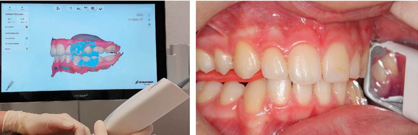
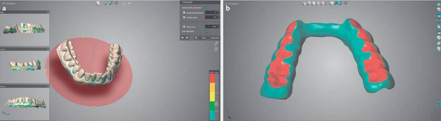
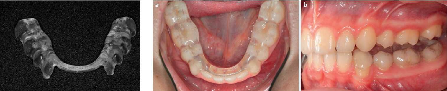
Occlusal splints are increasingly fabricated digitally today. First, the upper and lower jaws are captured contactlessly in 3D with an intraoral scanner. Then the relieving jaw position is determined manually and recorded with a construction bite made of 3 mm hard wax; this bite is also scanned. The practitioner plans the splint on the computer: it should fit well but not too tightly, and at its thinnest point be at least 1.5 mm thick so it does not break. It is fabricated by 3D printing from biocompatible plastic.7 In a randomized study, 3D-printed splints relieved symptoms as effectively as conventionally fabricated ones (Qin et al. 2025).8 The publication recommends printing two splints so that immediate replacement is available in case of loss, damage, or breakage. Today we fabricate the splints in our in-house master dental laboratory; because the splint is planned and printed digitally, printing a spare splint presents no difficulty.
What does the splint phase involve for you?
After examination and scan, we fabricate the splint in our in-house master dental laboratory in the same building — between the scan in the treatment room and the finished splint, there are no postal routes, just a hallway. The splint is usually worn at night; whether it is also needed during the day initially is determined based on your findings. After about six weeks, we review together whether the symptoms are decreasing and whether the jaw position remains stable; the splint is adjusted if needed. Splint therapy is usually scheduled for three to six months. How long it takes in your case depends on the findings. Only when the relieving position is established do we plan the next step.
Clinical and instrumental functional analysis is a private service. An occlusal splint based on digital jaw joint measurement (DMD) is also private and costs approximately 1,500 to 3,000 euros at our practice. We document the exact scope in advance in a cost estimate; details are available under TMD treatment.
What comes after the splint?
Once the jaw joints are relieved and the healthy position is established, the actual tooth correction can begin, often with clear aligners.
Read more:
- After the splint: Follow-up Treatment After Initial Splint Therapy (professional publication)
- Planning tooth correction: The Virtual Articulator in Aligner Orthodontics (professional publication)
- Treatment example: TMD pain with digitally fabricated splint
- TMD treatment at our practice · Aligners
Sources:
Underlying professional publication: Schupp, W., Haubrich, J., & Funke, J. (2020). Behandlung mit Okklusionsschienen und deren Herstellung im digitalen Workflow. Informationen aus Orthodontie & Kieferorthopädie, 52(3), 227–231. https://doi.org/10.1055/a-1200-5934
- Okeson, J. P. (2015). Evolution of occlusion and temporomandibular disorder in orthodontics: Past, present, and future. American Journal of Orthodontics and Dentofacial Orthopedics, 147(5), S216–S223. https://doi.org/10.1016/j.ajodo.2015.02.007
- Boisserée, W., & Schupp, W. (Hrsg.). (2012). Kraniomandibuläres und muskuloskelettales System. Quintessenz.
- Schupp, W., & Funke, J. (2018). Continuing diagnostics and therapy of the temporomandibular and musculoskeletal system: The rest position of the temporomandibular joint (TMJ) and the therapeutic construction bite vs. the centric bite. Journal of Aligner Orthodontics, 2, 267–281.
- Meyer, G. (2018). Short clinical screening procedure for initial diagnosis of temporomandibular disorders. Journal of Aligner Orthodontics, 2, 91–98.
- Schupp, W., Funke, J., Boisserée, W., et al. (2018). Continuing diagnostics of the temporomandibular and musculoskeletal system (TMS/MSS). Journal of Aligner Orthodontics, 2, 199–213.
- Al-Moraissi, E. A., Farea, R., Qasem, K. A., Al-Wadeai, M. S., Al-Sabahi, M. E., & Al-Iryani, G. M. (2020). Effectiveness of occlusal splint therapy in the management of temporomandibular disorders: Network meta-analysis of randomized controlled trials. International Journal of Oral and Maxillofacial Surgery, 49(8), 1042–1056. https://doi.org/10.1016/j.ijom.2020.01.004
- Lin, R., Yu, C. H., & Sun, J. (2019). Mechanical properties and cytotoxicity of three-dimensional printing polycarbonate for occlusal splints. Shanghai Kou Qiang Yi Xue, 28, 467–471.
- Qin, H., Liu, Y., Miao, H., et al. (2025). Comparative efficacy of digital 3D-printed and conventional stable occlusal splints in the treatment of temporomandibular disorders. Journal of Oral Rehabilitation, 52(10), 1699–1706. https://doi.org/10.1111/joor.14032
Your orthodontist in Cologne!
Schedule your no-obligation consultation at JUST KFO – Dr. Steinmaier & Kollegen in Cologne.
